
| 培养基名称: | SC-URA培养基 ,SC-U培养基,酵母SC-URA培养基、酵母SC-U培养基、酵母尿嘧啶缺陷型培养基;酵母尿嘧啶缺陷培养基、尿嘧啶缺陷筛选培养基 |
|---|---|
| 英文名称: |
SC-Dropout Medium SC-U Medium SC Dropout Medium without uracil Yeast Synthetic Drop-out Media |
| 产品目录号: | M300-01、M300-02、M300-08 |
| 产品规格: | 多规格 |
| 保存条件: | 密封,阴凉干燥处保存。 |
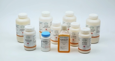
| 产品性状: |
浅黄色粉末。 液体浅琥珀色,透明,无沉淀或有轻微沉淀(不溶性硫酸钙)。 |
| 注意事项: | 避免摄入、呼入、皮肤接触。配制时在通风橱中进行,戴口罩、手套、护目镜。 |
| 相关产品: |
酵母氮源基础(YNB) SC-URA 氨基酸 酵母SC-Dropout氨基酸 |
酵母SC-URA培养基产品描述:
酵母SC-URA培养基又称酵母SC-U培养基,是合成和筛选培养基,用于筛选尿嘧啶营养缺陷型的酿酒酵母,是尿嘧啶营养缺陷型培养基。
SC-URA培养基与酵母SC完全(SC Complete medium)培养基相比,缺少尿嘧啶。不能合成尿嘧啶的酵母菌株在SC-URA dropout培养基中不能生长。
本产品中不含糖类,使用者可以根据实验目的添加葡萄糖、棉籽糖或半乳糖。
SC-URA尿嘧啶缺陷型酵母培养基用途
酵母SC-URA培养基用于筛选尿嘧啶营养缺陷型的酿酒酵母。
酵母SC-URA培养基添加半乳糖后,可作为酿酒酵母蛋白表达培养基。
酵母SC-URA培养基添加葡萄糖,可作为尿嘧啶缺陷型酵母传代和增菌用培养基。
由于SC-U培养基成分已知,因此可以用来鉴定酿酒酵母URA3营养缺陷型。
SC-Ura酵母培养基配方与配制方法
| 成分 | g/L |
|---|---|
| 酵母基础氮源(YNB) | 1.7 |
| 硫酸铵 | 5 |
| L-精氨酸 | 0.1 |
| L-半胱氨酸 | 0.1 |
| L-赖氨酸 | 0.1 |
| L-苏氨酸 | 0.1 |
| L-天冬酰胺 | 0.05 |
| L-异亮氨酸 | 0.05 |
| L-苯丙氨酸 | 0.05 |
| L-脯氨酸 | 0.05 |
| L-丝氨酸 | 0.05 |
| L-酪氨酸 | 0.05 |
| L-缬氨酸 | 0.05 |
| L-甲硫氨酸 | 0.05 |
| L-色氨酸 | 0.1 |
| L-组氨酸 | 0.1 |
| L-亮氨酸 | 0.1 |
| 腺嘌呤 | 0.1 |
| YNB成分 | mg/L |
|---|---|
| 肌醇 | 2.0 |
| 烟酸(维生素B3) | 0.4 |
| 盐酸硫胺素(维生素B1) | 0.4 |
| 硫酸铜 | 0.04 |
| 磷酸二氢钾 | 1000 |
| 硼酸 | 0.5 |
| 盐酸吡哆醇(维生素B6) | 0.4 |
| 泛酸钙(维生素B5) | 0.4 |
| 对氨基苯甲酸 | 0.2 |
| 硫酸镁 | 500 |
| 硫酸锰 | 0.4 |
| 硫酸锌 | 0.4 |
| 氯化铁 | 0.2 |
| 核黄素(维生素B2) | 0.2 |
| 氯化钙 | 100/td> |
| 碘化钾 | 0.1 |
| 钼酸钠 | 0.2 |
| 生物素(维生素B7、H) | 0.002 |
| 氯化钠 | 100 |
配制方法:

1. 在分析天平上称取SC-U培养基7.9g,加900 mL去离子水溶解。

2. 15 psi, 121°C 灭菌15 min。

3. 冷却至50°C左右,加入100 mL的20%葡萄糖溶液(预先配制,用0.2微米滤膜过滤除菌),根据实验需要,也可以用棉籽糖、半乳糖。
乳糖诱导的酿酒酵母蛋白表达实验流程
GAL1 Promoter
In INVSc1, transcription from the GAL1 promoter is repressed in the presence of glucose (West et al., 1984). Removing glucose and adding galactose as a carbon source induces transcription (Giniger et al., 1985). Maintaining cells in glucose gives the most complete repression and the lowest basal transcription of the GAL1 promoter. Transferring cells from glucose- to galactose-containing medium causes the GAL1 promoter to become de-repressed and allows transcription to be induced.
Alternatively, cells may be maintained in medium containing raffinose as a carbon source. The presence of raffinose does not repress or induce transcription from the GAL1 promoter. Addition of galactose to the medium induces transcription from the GAL1 promoter even in the presence of raffinose. Induction of the GAL1 promoter by galactose is more rapid in cells maintained in raffinose when compared to those maintained in glucose.
You may choose to grow cells containing your pYES fusion vector in glucose or raffinose depending on how quickly you want to obtain your expressed protein after induction with galactose and on the toxicity of the expressed protein. For more information about expression in yeast, please refer to the Guide to Yeast Genetics and Molecular Biology (Guthrie and Fink, 1991).
诱导实验
To induce expression of your protein of interest from the GAL1 promoter, galactose is added to the medium. For cells that have been maintained in glucose, recombinant fusion protein can be detected in as little as 4 hours after galactose induction. Recombinant fusion protein can be detected in cells that have been cultured in raffinose by 2 hours after galactose induction.
If you are assaying for expression of your recombinant fusion protein for the first time, we recommend that you perform a time course to optimize expression of your recombinant protein (e.g. 0, 4, 8, 12, 16, 24 hours after galactose induction). A standard protocol is provided below to perform a time course experiment. Other protocols are suitable.
1. Inoculate a single colony of INVSc1 containing your pYES fusion vector into 15 ml of SC-U selective medium containing 2% glucose or 2% raffinose. Grow overnight at 30°C with shaking.
2. Determine the OD600 of your overnight culture. Calculate the amount of overnight culture necessary to obtain an OD600 of 0.4 in 50 ml of induction medium (SC-U selective medium containing 2% galactose).
Example: Assume that the OD600 of an overnight culture is 3 OD600 per ml. Then, the amount of overnight culture needed to inoculate a 50 ml culture to OD600 = 0.4 is (0.4 OD/ml) (50 ml) = 6.67 ml 3 OD/ml
3. Remove the amount of overnight culture as determined in Step 2 and pellet the cells at 1500 x g for 5 minutes at room temperature. Discard the supernatant.
4. Resuspend the cells in 50 ml of induction medium. Grow at 30°C with shaking.
5. Harvest an aliquot of cells at 0, 4, 8, 12, 16, and 24 hours after addition of cells to the induction medium. For each time point, remove 5 ml of culture from the flask and determine the OD600 of each sample. You will use this information when assaying for your recombinant fusion protein.
6. Centrifuge the cells at 1500 x g for 5 minutes at +4°C.
7. Decant the supernatant. Resuspend cells in 500 µl of sterile water.
8. Transfer cells to a sterile microcentrifuge tube. Centrifuge samples for 30 seconds at top speed in the microcentrifuge.
9. Remove the supernatant.
10. Store the cell pellets at -80°C until ready to use. Proceed to the next section to prepare cell lysates to detect your recombinant protein.
检测目的蛋白
Detection of To detect expression of your recombinant fusion protein by western blot, you Recombinant may use the Anti-V5 or the Anti-His antibodies or an antibody to your protein of interest.
You will also need to prepare a cell lysate from your yeast transformant. A general protocol for small-scale preparation of cell lysates using acid-washed glass beads is provided below for your convenience. Other protocols are suitable. Please refer to Current Protocols in Molecular Biology, Unit 13.13 (Ausubel et al., 1994) for more information. For large-scale preparations (culture volumes over 1 liter).
Breaking buffer (50 mM sodium phosphate, pH 7.4, 1 mM EDTA, 5% glycerol, 1 mM PMSF).
Acid-washed glass beads (0.4-0.6 mm size; Sigma-Aldrich, Catalog no. G8772).
1. You may prepare cell lysates from either frozen cells or fresh cells.
Reminder: You will need to know the OD600 of your cell sample(s) before beginning (see Step 5).
2. Resuspend fresh or frozen cell pellets in 500 µl of breaking buffer. Centrifuge at 1500 x g for 5 minutes at +4°C to pellet cells.
3. Remove supernatant and resuspend the cells in a volume of breaking buffer to obtain an OD600 of 50-100. Use the OD600 determined in Step 5, previous page, to calculate the appropriate volume of breaking buffer to use.
4. Add an equal volume of acid-washed glass beads.
5. Vortex mixture for 30 seconds, followed by 30 seconds on ice. Repeat four times for a total of four minutes to lyse the cells. Cells will be lysed by shear force. You can check for the extent of lysis by checking a small aliquot under the microscope.
6. Centrifuge in a microcentrifuge for 10 minutes at maximum speed.
7. Remove supernatant and transfer to a fresh microcentrifuge tube. Assay the lysate for protein concentration using BSA as a standard.
8. Add SDS-PAGE sample buffer to a final concentration of 1X and heat the sample for 5 minutes at 70°C.
9. Load 20 µg of lysate onto an SDS-PAGE gel and electrophorese. Use the appropriate percentage of acrylamide to resolve your recombinant protein.
放大实验
Once you have determined the optimal induction time necessary to obtain maximal protein expression, you may increase the protein yield by scaling up the procedure. If you plan to use ProBond resin to purify your recombinant fusion protein, please see the Note below. To prepare cell lysates from culture volumes over 1 liter, we recommend that you use a bead beater to lyse the cells. Please refer to Current Protocols in Molecular Biology, Unit 13.13 (Ausubel et al., 1994) for a suitable protocol to lyse cells with a bead beater.

